Advancements In Imaging For Endometriosis, Diagnostic Care And Impact On Healthcare In Canada
The Endometriosis Network Canada (TENC) met with endometriosis expert Dr. Mathew Leonardi to discuss advancements in imaging for endometriosis and diagnostic care and its potentially life-altering implications for patients for Endometriosis Awareness Month 2021. The conversation continues as Dr. Leonardi provides insights for fellow physicians and an overview about the role imaging plays to the benefit of the healthcare system in Canada.
Dr Mathew Leonardi is an expert in complex gynecology, endometriosis excision surgery and gynaecological ultrasound (associate professor) at McMaster University Medical Centre in Hamilton, Canada. He is an honorary adjunct lecturer at the University of Adelaide. His philosophy of care includes working in an interdisciplinary team and patient-centred decision making. He has been awarded his PhD from the University of Sydney which is focused on the utility of ultrasound in the diagnosis and surgical management of endometriosis.
Dr Leonardi is a nationally and internationally recognized leader in his field. He has published over 120 peer-reviewed scientific articles, several textbook chapters, and presented at numerous international congresses on endometriosis. He has received numerous awards for his conference presentations on endometriosis. He is an avid researcher, actively contributing academically to the advancement of gynecologic health.
Dr. Leonardi is on the World Endometriosis Society Early Career Board and a founding member of the Next Generation Committee at the International Society of Ultrasound in Obstetrics and Gynecology. He is an Associate Editor for Reproduction & Fertility and on the Editorial Board for the Journal of Minimally Invasive Gynecology, Ultrasound in Obstetrics and Gynecology, and the Journal of Obstetrics and Gynaecology of Canada. He is part of an international research group named Imagendo, which won the most prestigious science award in Australia, the 2023 ANSTO Eureka Prize for Innovative Use of Technology.
He believes in listening to patients, validating their experiences, and working together with the patient to achieve the best outcomes.
The Endometriosis Network Canada (TENC): What are some ways imaging can be used to help physicians manage endometriosis in patients?
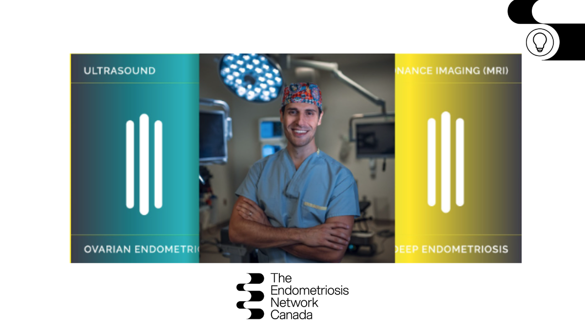
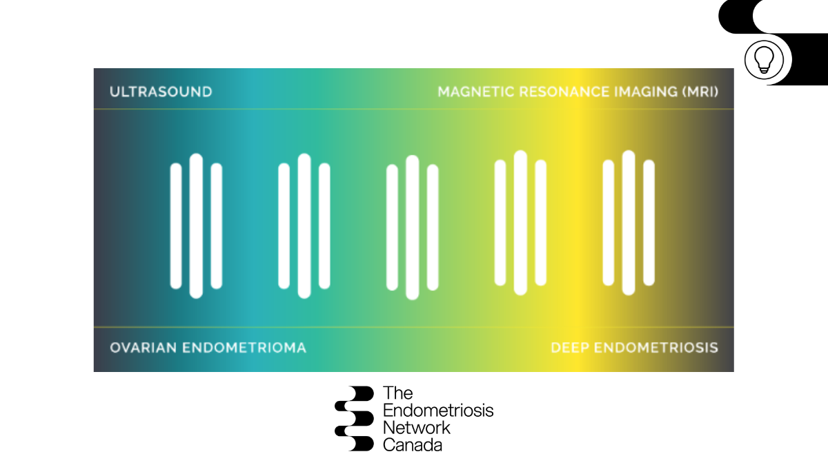
Dr. Mathew Leonardi (ML): There are two broad imaging modalities currently used to assist in diagnosing endometriosis: ultrasound and magnetic resonance imaging (MRI). Imaging can help us understand and map out disease sites. Knowing where those lesions are very helpful for surgeons, gynaecologists, even family doctors, when speaking to patients about their options for management of the disease.
Through imaging, physicians can provide a visual diagnosis (i.e. radiologic diagnosis) for a subset of patients. This includes individuals with ovarian endometriomas, deep endometriosis, and severe pelvic adhesions. At present, the gold standard of diagnosis of endometriosis is surgery, which to me – and maybe to some of you as well – is kind of puzzling because surgery is invasive and comes with its own risks.
From a medical professional standpoint, I believe that by moving to a system where there are multiple avenues to diagnose patients, including imaging based or radiologic, it would minimize risks associated with surgery to the ultimate benefit of patients and the healthcare system. While we could then avoid the surgical diagnosis in this specific group of people, it is not possible to do this in every case – yet.
We can also start to see more disease through imaging, which means we can now validate some patients with a visual radiologic diagnosis. And I think this is really important because when patients have experienced symptoms for a long time and are told repeatedly that “things are normal” and “all in their head,” it leads to worsened outcomes.
TENC: The implications of this on the health and well-being of patients could be life-changing.
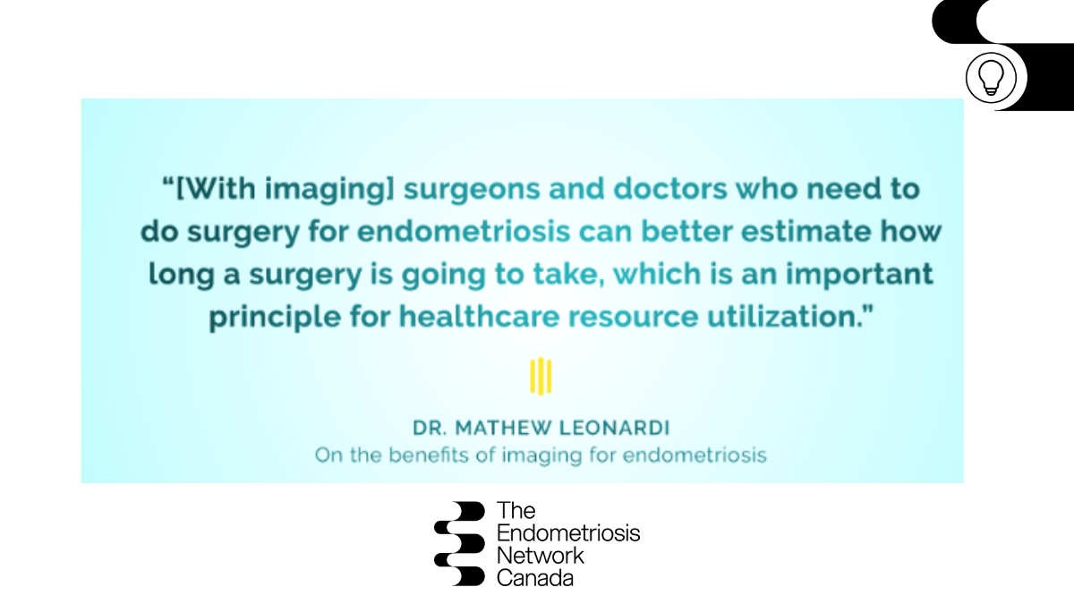
ML: Without a doubt! Additionally, if you as a gynaecologist are considering surgery as a treatment option, imaging equips you with the knowledge to know whether other experts are needed to operate with you. For example, if the patient warrants bowel surgery for endometriosis, we may have to ask our colorectal surgeons to come with us. If there is a large bladder nodule present, we have to be prepared to bring our urology colleagues with us.
Knowing about these in advance not only prepares the medical team, it importantly allows for patients to meet the surgeon, have a proper consent process, and prepare for the surgery.
We also have the opportunity to shift our focus exclusively from treatment discussions to diagnosis as the presence of imaging tests increases across Canada.
“We need endometriosis experts who are experts in the diagnosis and treatment of the disease.”
TENC: How can physicians facilitate a proper consent process with their patients with the help of imaging?
ML: I believe this comes down to the capacity of imaging to help establish an informed consent process. When we know more and have more to share about the disease then go to talk to patients about their options – medical, surgical management, multidisciplinary management with physiotherapy, psychotherapy, chronic pain team management, the patient has a better opportunity to provide targeted, informed consent. This is in contrast to the current climate where I believe patients are generally told ‘we think you have endometriosis; this is what we should do for you.’ Because there is not a refined understanding of the disease in such situations, patients are not able to make a fully informed decision, and therefore, informed consent.
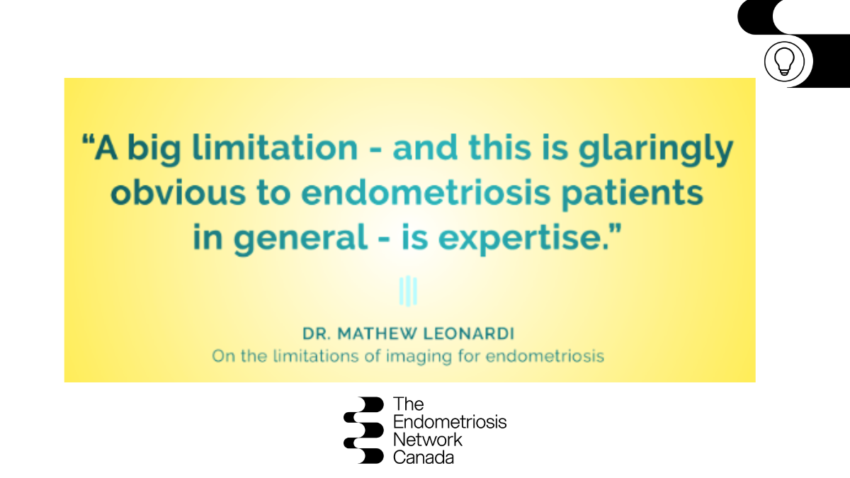
TENC: We spoke in Part I about the limitations with imaging and you mentioned a gap in expertise. How can the healthcare system address this?
ML: Furthering education and fostering interest in this specialized field among medical students and established professionals. This is why I’m training more fellows in our fellowship program and what I’m educating people about by speaking at opportunities like this, in addition to as many conferences, educational webinars and sessions as possible. This is what we as experts need to do to overcome this existing limitation surrounding education and training.
TENC: What are the advantages of using imaging to diagnose endometriosis?
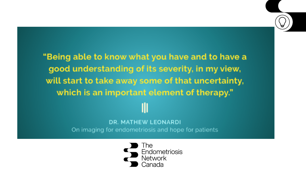
ML: We can assess the severity of disease. When I am scanning patients in the office, and we see deep endometriosis, we see adhesions, we can give them an important understanding of the severity. We are also able to tell them that if we do not see endometriosis on the scan, it doesn’t mean that there is no endometriosis present. But we at least know we are not dealing with severe endometriosis because severe disease is especially easy to see on ultrasound.
Ultrasound and MRI are also much more affordable than surgery. They are very safe, and in my experience, patients generally find them acceptable and approachable diagnostic tools.
Imaging provides a totally new and different way of looking at the pelvis. I know from spending a lot of time on social media groups that there is this mentality that sometimes surgeons miss endometriosis at surgery. That could be because they may not be trained in the area of endometriosis or that the adhesion state is so severe that it’s very difficult to tell what is actually within the adhesions themselves that’s causing the adhesions. Ultrasounds can see through such things. MRIs can similarly see through things that we cannot see with just our eyes at surgery.
“One of my strategies is to engage with the endometriosis community directly because for a long time the community has driven change.”
TENC: What is one action fellow medical professionals can take today to optimize care for patients when it comes to imaging?
ML: If you are a gynaecologist that works somewhere near the Hamilton area, please reach out. Send me imaging referrals for your patients and I’ll provide you with a very detailed report that you can then review with your patient and discuss their ongoing management.
In fact, several doctors in the region are already doing this, which is amazing. I find that patients are grateful for the opportunity, even if they must travel a little bit for the test. The same would apply if you’re working in British Columbia or in the Ottawa area, because those tools exist. You just have to work a little harder to access them.
TENC: Dr. Leonardi, we cannot thank you enough for sharing your expertise, for educating both patients and physicians, and ultimately, for inspiring hope.
ML: I am so honoured to be here to work with all of you to improve the state of endometriosis care in Canada and internationally as well.
Learn more about Dr. Leonardi, his research, work, and practice at: mathewleonardi.com
Connect with him on social:
Instagram – @drmathewleonardi
Twitter – @MathewLeonardi
Facebook – @drmathewleonardi
Stay tuned for the full-length video interview with more information for patients and clinicians — coming soon!
Read Part I of the ‘Imaging for Endometriosis’ interview with Dr. Leonardi